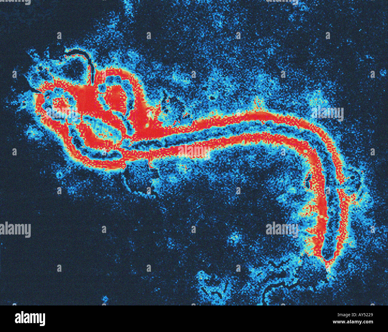
Virus Ebola Microscope / Ebola Virus Microscope High Resolution Stock Photography And Images Alamy - Fluorescent microscope image showing an ebola strain growing in cells in a cdc lab.
Virus Ebola Microscope / Ebola Virus Microscope High Resolution Stock Photography And Images Alamy - Fluorescent microscope image showing an ebola strain growing in cells in a cdc lab.. Diese pandemie kam nicht zufällig zustande. In february 2014, piot returned to yambuku for only the second time since 1976, to mark his 65th birthday. Cdc / fred a murphy). In particular, the generic term ebola virus is widely used to refer specifically to members of the species zaire ebolavirus. After entering the body, it kills cells, making some of them explode.
13, 1976, exactly 38 years ago monday, frederick a. Understanding the ebola virus like all viruses, an understanding of the unique structure is critical to the pursuit of successful therapies. Ebola is a deadly disease caused by a virus. An electron micrograph (25,000x magnification) of ebola virus particles (green) attached to, and budding from, an infected cell (blue). Unlike hiv, ebola is not an unknown virus.
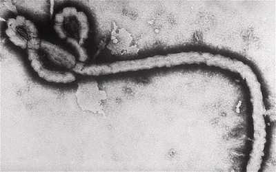
Scanning Electron Microscope Sem Image Of The Ebola Virus Stock Photo Alamy from c8.alamy.com Yet the coronavirus genome remains a mystery, especially to those outside the scientific community. Consequently, in 2010, a group of researchers recommended that the name ebola virus be adopted for a subclassification note 1 within the species zaire ebolavirus and that similar common names be formally adopted for. Transmission electron microscopy of the ebola virus daniel r. In february 2014, piot returned to yambuku for only the second time since 1976, to mark his 65th birthday. Scanning electron microscope image of the filamentous ebola viruses (stained blue) in an infected cell. People can get evd through direct contact with an infected animal (bat or nonhuman primate) or a sick or dead person infected with ebola virus. After multiplying inside a host cell, the stringlike ebola virus is emerging to infect more cells. The ebola river in 1976.
The ebola virus has a distinct elongated shape— a popular electron microscope image taken by frederick murphey resembled a long shepherd's crook.
Unlike hiv, ebola is not an unknown virus. Find professional ebola virus microscope videos and stock footage available for license in film, television, advertising and corporate uses. Murphy appears in the hot zone and his now famous photo of the ebola virus appears in the film outbreak. Murphy is considered one of the world authorities on viruses. The tests detected the virus from. A few people are still infected with ebola each week. An outbreak of a wildly deadly virus on the doorstep of the nation's. Booth national microbiology laboratory, 1015 arlington street, winnipeg, manitoba, r3e 3r2, canada. Previously, the virus was identified in blood samples using an electron microscope. Consequently, in 2010, a group of researchers recommended that the name ebola virus be adopted for a subclassification note 1 within the species zaire ebolavirus and that similar common names be formally adopted for. Price batelle press, columbus, ohio isbn: Images showing individual symptoms of ebola. Goldsmith was able to take a different one that looks like a long thin filamentous particle, which was featured widely in publications during the 2014 ebola outbreak.
Murphy had been there when ebola was named in 1976; Previously, the virus was identified in blood samples using an electron microscope. Murphy appears in the hot zone and his now famous photo of the ebola virus appears in the film outbreak. The ebola river in 1976. October 13, 2014 4:15 pm edt o n oct.
Ebola Virus Disease Pictures Symptoms Victims And Relief Work Enkivillage from i.enkivillage.org Murphy had been there when ebola was named in 1976; Find professional ebola virus microscope videos and stock footage available for license in film, television, advertising and corporate uses. Transmission electron microscopy of the ebola virus daniel r. Ebola is a deadly disease caused by a virus. After entering the body, it kills cells, making some of them explode. Under an electron microscope, it looks like a harmless shepherd's crook or a scheerio with a long tail, but it can decimate the human immune system. Vesicular stomatitis virus (vsv) under an electron microscope (original image: He met sukato mandzomba, one of the few who.
Murphy, a cdc virologist and expert in photographing viruses peered into a microscope and saw what he.
There are five strains, and four of them can make people sick. October 13, 2014 4:15 pm edt o n oct. A virus is a submicroscopic infectious agent that replicates only inside the living cells of an organism. The cdc has been dealing with ebola since the first case was discovered in africa in 1976. Vesicular stomatitis virus (vsv) under an electron microscope (original image: Ebola virus disease (evd) is a rare and deadly disease in people and nonhuman primates. A view of infection using electron microscopy elena i. In particular, the generic term ebola virus is widely used to refer specifically to members of the species zaire ebolavirus. Ebola is a deadly disease caused by a virus. Transmission electron microscopy of the ebola virus daniel r. Viruses infect all life forms, from animals and plants to microorganisms, including bacteria and archaea. Under an electron microscope, it looks like a harmless shepherd's crook or a scheerio with a long tail, but it can decimate the human immune system. The tests detected the virus from.
Transmission electron microscopy of the ebola virus daniel r. Murphy had been there when ebola was named in 1976; Green cells are those infected with ebola and red are uninfected cells. Here's the coronavirus under an electron microscope. There are five strains, and four of them can make people sick.
Transmission Electron Microscope View Of An Ebolavirus Virion The Bar Download Scientific Diagram from www.researchgate.net Vesicular stomatitis virus (vsv) under an electron microscope (original image: The virus is very resilient, killing the virus requires high doses of gamma irradiation and ultraviolet light, long periods of 30 minutes or more of intense heat 60 degrees celsius (140 degrees fahrenheit). Price batelle press, columbus, ohio isbn: Booth national microbiology laboratory, 1015 arlington street, winnipeg, manitoba, r3e 3r2, canada. Scanning electron microscope image of the filamentous ebola viruses (stained blue) in an infected cell. People can get evd through direct contact with an infected animal (bat or nonhuman primate) or a sick or dead person infected with ebola virus. An electron micrograph (25,000x magnification) of ebola virus particles (green) attached to, and budding from, an infected cell (blue). Recognise and control new variants of the deadly ebola virus more quickly.
A view of infection using electron microscopy elena i.
But he had no way of. A view of infection using electron microscopy elena i. Booth national microbiology laboratory, 1015 arlington street, winnipeg, manitoba, r3e 3r2, canada. October 13, 2014 4:15 pm edt o n oct. Understanding the ebola virus like all viruses, an understanding of the unique structure is critical to the pursuit of successful therapies. Previously, the virus was identified in blood samples using an electron microscope. When the scientists, inside their belgian laboratory, looked under the microscope at blood samples sent from africa, the virus looked like a worm or a long string, unlike almost any viruses known. Price batelle press, columbus, ohio isbn: Yet the coronavirus genome remains a mystery, especially to those outside the scientific community. A virus is a submicroscopic infectious agent that replicates only inside the living cells of an organism. A few people are still infected with ebola each week. Fluorescent microscope image showing an ebola strain growing in cells in a cdc lab. The ebola virus has a distinct elongated shape— a popular electron microscope image taken by frederick murphey resembled a long shepherd's crook.
He also helped discover the marburg virus, a sister to ebola zaire virus ebola. Murphy is considered one of the world authorities on viruses.


0 Komentar